A Heartwarming Tale Of Orphaned Girls Finding a Home

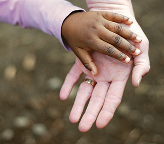
When the Asian tsunami of 2003 hit India, hundreds of thousands of lives were lost, and hundreds of children were orphaned. Shortly after the tragedy, a woman based in the United States named Sumathi thought about adopting an orphan or two. However, she also considered her age, and so she thought it best to build a home for them in her native country of India.

44-830x268.jpg
Image source: college4u.in

It wasn’t easy since the legalities of adopting that many children take a lot of time and effort. After years of orphanages being used as fronts for farming children for their organs and as prostitution dens, the government wasn’t going to make it easy.

But through perseverance, Chinmaya Vijaya Orphanage opened its doors to children who no longer had people to take care of them. It took 18 months to open the 13-employee house, but with a monthly pledge of $2,000 from benefactors and other kind sponsors, the first 20 girls were taken from a WHO shelter and given a home. These girls were indeed rescued from slums and “guardians” who had them do heavy manual labor for cash.

Not waiting for them to be used as prostitutes, Sumathi made her move and took in the girls. Today, the number is up to 50 girls between ages four and eight. The target of the orphanage is to adopt 200.

hqdefault
Image source: youtube.com

Dr. Apparao Mukkamala is a clinical professor of radiology at the Michigan State University College of Human Medicine and was among the founders of Chinmaya Vijaya Orphanage. Learn more about his work here.

Opening New Horizons For Radiology Through Artificial Intelligence

Machine learning and artificial intelligence (AI) systems are commonplace in many industries today. Progress has been swift in the development of AI systems in medical fields such as radiology. Meanwhile, researchers from New York’s Icahn School of Medicine at Mount Sinai have developed machine learning technology that could interpret radiologist reports with an accuracy rate of 91 percent. In another example, swarm AI systems allowed groups of radiologists to reduce false positives and false negatives without having to meet to cross-examine data.

Image source: contentful.com

Some of the possible applications of machine learning and other AI developments in radiology include identifying cases that require immediate evaluation, highlighting anomalous results of cross-sectional imaging for further review, and characterizing masses for malignancy. The increase in diagnostic accuracy could in turn help doctors identify problematic areas early and recommend more effective interventions and care to patients.

Machine learning and AI systems would also work brilliantly alongside image sharing networks, gleaning and learning from a diverse array of information from patients to further increase their accuracy.

Image source: sundaypost.com

AI and machine learning systems in radiology are still largely in their infancy. Limitations, such as the lack of sufficient data from which the systems could learn, are still a cause for concern for many experts. In addition, AI systems cannot always account for variations with a population. Some people without any anomaly, for instance, would fall out of the statistical baseline on which an AI relies.

Dr. Apparao Mukkamala served as a clinical professor of Radiology at the Michigan State University College of Human Medicine and was a board member and chair of the Department of Radiology at Hurley Medical Center. Visit this blog for more updates on the innovations surrounding radiology.

Brilliant Charitable Event Ideas For Children

One of life’s greatest joys is seeing children happy. This is why one of philanthropists’ favorite events to organize are fundraisers for children who are either sick and confined to the hospital or in an orphanage.

6278fb8356bceece76aff1ae57b299214ed674396d87f4.23678124
Image source:www.smartbusinessdirectory.co.uk

While standard fundraisers with food and drinks and maybe a show may seem like a good idea, there are other really wonderful alternatives that not only adults but the kids as well will get a blast out of. And if it’s a fundraiser, perhaps attendees will see the value in the activity and may want to contribute even more than they initially planned.

Below are a few ideas that are sure to get the kids’ attention.

Petting Zoo

Kids always love cute, cuddly animals. Having dogs, squirrels, lambs, and small horses in the area seems like a fun afternoon. Thrown in a horseback ride and an animal show, and it’ll be a day the kids will never forget.

Go Karts

What kid doesn’t love little cars? Parties in Go Kart parks are always a hit among children. Having a fundraiser in such a place can be a blast for both children and adults in attendance.

goat feeding
Image source:eckerts.com

Boat Ride

At first glance, boat rides may seem boring. But with a child’s imagination, and with wildlife in the area, it can be an adventure they’d want to do over and over again.

Dr. Apparao Mukkamalais a radiologist currently based in Flint, MI. Together with his wife, Dr. Sumathi R. Mukkamala, he helped establish the Chinmaya Vijaya Orphanage, which delivers care and education to orphaned and abandoned girls throughout India. Learn more about his work by checking out this blog.


A Closer Look At The Changing Landscape Of Radiology

Image source: medicalfuturist.com

Technology continues to be the main force driving advancements in radiology. While some innovations are seen to play a bigger role in the coming years, artificial intelligence and 3D printing have already made a huge impact in the medical profession in 2017.

3D printing, in particular, has given radiologists and other clinicians the opportunity to realize studies previously only available in CT, 3D, and MRI modalities. Companies are developing ways to generate anatomical models from 3D output files, effectively changing the way we look at clinical education and patient engagement.

Collaborations are also becoming common among pharmaceutical and medical research companies and web apps. For example, we now have the app Teledoc in the Apple store or in Google Play, which claims that it can connect users to 3,600 licensed healthcare professionals via video or phone call, with an average doctor response time of 10 minutes, 24 hours a day.

Image source: siemens.com

Wearables and the Internet of Things (IoT) should become more common in diagnostic imaging by 2018. A number of applications have emerged to push IoT into diagnostic imaging, including remote inventory monitoring and health monitoring, controlling energy use, and monitoring engine performance.

Finally, as far as wearables are concerned, we’ve seen the rise of smartwatches in 2017, which bring health and fitness apps directly to our wrists. Among these are the Apple watch and Pebble.

Based in Flint, MI, Dr. Apparao Mukkamala is a clinical professor of radiology at the Michigan State University College of Human Medicine. He also served as a board member and chair of the Department of Radiology at Hurley Medical Center. For more on his work, visit this website.

Recent breakthroughs in medical care

While 2016 was an excellent year for medicine and medical technology in general, with the rise of virtual and augmented reality and smarter algorithms for analyzing data, 2017 has been even more promising for the industry. Here are some breakthroughs that have been further revitalizing the field of medical care.

2-lab.jpg

Image source: medicalxpress.com

The famed IBM Watson advanced artificial intelligence program is one of the most significant devices transforming health care. This computer has allowed physicians to quickly go through streamlined personal reports of patients, instead of settling for tons of individual papers per patient. The Watson has been particularly useful in both oncology and radiology (for the latter, in the Medical Sieve project), with the specialized Watson for Genomics project doing revolutionary work in advanced precision medicine.

Oncology has greatly benefited from precision medicine this year, with digital technology changing the way doctors deal with available cancer care methods. Personalized oncology is now available through startups and companies such as Foundation Medicine and SmartPatients, allowing for patient empowerment online. Immunotherapy in the form of liquid biopsy started gaining traction in 2016, and it’s now becoming more accessible to patients all over the globe.

Finally, 2017 has ushered in a new era in diabetes care and management with the United States’ FDA approval of the world’s first artificial pancreas. This landmark device automatically monitors blood sugar and supplies insulin, replicating what the organ does on its own. It has enabled diabetes patients to live a more convenient, sustainable, and easier life.

IBM-Watson-in-Oncology

Image source: medicalfuturist.com

Dr. Apparao Mukkamala is the former president of the Michigan State Medical Society. Today, he is a clinical professor of radiology at the Michigan State University College of Human Medicine. For more reads on the field of medicine, drop by this blog.

A passage to India: US-based radiologist gives back to the orphans of his native land

Radiologist Dr. Apparao Mukkamala, now based in Flint, Michigan, was flipping through some photos of orphaned and abandoned girls whom he and his wife has helped years back. These young women mostly come from poor, rural villages of the southeastern coast of India. They used to live in shanties or on the streets, beggars with no care for their looks and health. Parasites lived on their hair and bodies. A lifetime of either prostitution or slavery awaited them.

3368317171_532ae24d77_z
Image source: flickr.com

Now, Dr. Apparao can’t help but smile, seeing the very same girls in uniforms, arms draped around each other in a gleeful air. They are surrounded by comfort and are in pursuit of learning: they have books, they write, play the piano. They now dream of being doctors themselves, empowered to lead in service.

“The only limit they have now is their dreams,” says Mukkamala, beaming with pride. “They didn’t even know the alphabet before. It is so gratifying to see them full of ambition, smiling and making their future.”

The good doctor has been granted multiple awards in India for his charitable work and large-scale humanitarian efforts, including the Distinguished Citizen Award from the Tall Pine Council-Boy Scouts of America.

Nowadays, Apparao Mukkamala spends nearly 12 weeks of the year in their native state of Andhra Pradesh. His wife Dr. Sumathi Mukkamala, a pediatrician, spends more than six months there. There are now over a hundred girls living in their orphanage Chinmaya Vijaya, and the children call them “mommy” and “daddy.”

“I’m humbled,” says Mukkamala, who is blessed with two children and four grandchildren. “Happiness is in giving, not taking.”

maxresdefault
Image source: youtube.com

Radiologist Dr. Apparao Mukkamala is currently based in Flint, MI. He and his wife Dr. Sumathi R. Mukkamala helped establish the Chinmaya Vijaya Orphanage, which delivers care and education to orphaned and abandoned girls throughout India. More on his advocacies and work here.

The early history of radiology

Although X-rays were discovered by Wilhelm Roentgen, radiology as a field can owe its existence to Paul Hodges. Hodges was a bright young fellow who was raised by his uncle, Dr. William Rinehart after the young boy’s father passed away. It was also a fortunate coincidence that Dr. Rinehart worked closely with Roentgen during those days.

T_0217_x-ray_464753194_A
                                                                       Image source: nhs.uk

In 1897, radiology in its earliest form was started in Rinehart Hospital. The first X-ray machine had an induction coil, a mechanical interrupter, and a small X-ray tube, like the one used by Roentgen. Young Paul Hodges saw this machine as a kid. By the time he reached his early teens, Hodges became an apprentice at the hospital where he would have access to more sophisticated X-ray equipment. He also practiced preparing chemical solutions that were used in X-ray photography.

After Hodges got his M.D. at Washington University in 1918, he began teaching physiology to nurses. He had a recording device that a quartz fiber galvanometer, and a Brown tube (which could show electron streams). Hodges would work closely with other researches on animals.

That same year, Hodges would become a commissioned officer in the U.S. Army. Again, through fortunate coincidence, his expertise brought him to X-ray School in Camp Greenleaf, Georgia. It was his creation of a research department at the University of Chicago many years later that brought radiology to a whole different level.

surgery-radius-technology-x-ray-xray_1172-444
                                                                       Image source: freepik.com

Dr. Apparao Mukkamala is a clinical professor of Radiology at the Michigan State University College of Human Medicine. For more about his medical profession and his other work, visit this website.

A primer on voluntary employee beneficiary association

The existence of a voluntary employee beneficiary association or VEBA is a sign of a healthy, employee-mindful company. A VEBA exists for the sole purpose of promoting and championing a wide range of employee benefits– from accident insurance and legal services to vacation and severance pay, childcare, and life insurance benefits.

16687016354_ca450d18ec_z
                                                          Image source: managementink.wordpress.com

A VEBA is essentially a tax-exempt, in-house trust fund for everything related to employee concern. It cannot earn for the benefit of non-employee members; no private individual or group can benefit monetarily from work in a VEBA.

VEBAs must have no other function than the handling of employment-related matters, ensuring that members have access to all benefits covered by the collective bargaining agreement with the company. Also, as its name insists, VEBA’s membership must be purely voluntary.

In 2007, the United Auto Workers agreed to form VEBAs for its workers, a breakthrough decision that helped ease the mandatory health benefit burdens of their respective car company employers. As of March 2014, the UAW’s Retiree Medical Benefits Trust had more than $58.8 billion in assets, making it the world’s largest VEBA.

download
                                                               Image source: autonews.com

Dr. Apparao Mukkamalais a radiologist currently based in Flint, MI. In 2007, he became the president of the Michigan State Medical Society (MSMS). In 2008, he led a seminar in MSMS on the many benefits of the UAW VEBA that led to better health insurance benefits for member employees. For more on his work and advocacies, click here.

Beyond Adoption:Three Ways To Support An Orphanage

Helping orphaned and abandoned children goes beyond the noble act of adoption. While there are individuals who see adoption as the best option for them, there are also others who are willing to help in another way. Unfortunately, they are also unaware of the many ways they can support children living in orphanages. Below are some ways individuals can lend their resources to these homes:

08_Mavis2_JN
Image source:Christianstandard.com

Sponsor a child

One of the common problems orphanages face involves funding. Without enough financing, they won’t be able to give quality provision to the children under their care. Willing individuals can choose to sponsor a child, covering clothing, food, and even education for a year or more. Others can also donate to the orphanage, allowing them to divide the finances accordingly.

Be a short-term foster parent

Some programs allow those who are considering adoption to try first being a foster parent. For weeks up to a month, an individual, couple, or family can adopt a child who will stay in their home. This allows a child to experience the joy of being part of a family. For crowded orphanages, this allows the social workers focus on caring and giving attention to the children who have been left in the home.

Volunteer regularly

One of the problems orphanages face are the so-called “voluntourists” who travel overseas to witness what is happening to abandoned children in an impoverished country. While their help and visit are very much welcome, these people are not able to fully grasp the situation the institution is facing.They are also unable to provide consistent and timely help. A better way to support orphanages is by volunteering regularly. Being consistent in lending a hand for the children and social workers will help a person fully understand the situation and witness the developments.


pakistan-s-orphans-1463914740-1479
Image source:Nation.com.pk

Providing support to children in orphanages is a worthy cause.While there are many options available, an individual must be wise in choosing where and when he will invest his time, effort, and resources.

Dr. Apparao Mukkamala is the primary founder of NRI Medical Center and Medical School in Andhra Pradesh and, alongside his wife, Dr. Sumathi R. Mukkamala, the co-founder of Chinmaya Vijaya Orphanage. Visit this page for similar reads.

Everything a Patient Needs To Know About Ct Scans

Sometimes, a radiologist may recommend certain medical imaging procedures to gain a better understanding of a patient’s condition and how to best treat it. One of the more common tests prescribed is the computed tomography (CT) scan, otherwise known as the CAT scan.This test uses x-rays to project an image of different parts of the body and detect any abnormalities that may be present in soft tissue. This is the most “basic” test in that it cannot detect little variables within harder structures such as a tumor growth in a bone marrow. Despite this limitation, doctors typically request for a CT scan first to eliminate obvious potential damages. Listed below are a list of everything a patient needs to know about the procedure.

ct-scanner
Image Source: medicalnewstoday.com

It is an outpatient test: CT scans are done in a hospital or an outpatient facility.Scans are painless and take only a few minutes to conduct, especially among facilities that have newer machines. Patients can expect to take only 30 minutes for the entire procedure. CT scanners are usually shaped like a large donut standing on its side. Patients are requested to lie on a motorized table that slides through the opening of the tunnel. When the table moves into the scanner, x-ray tubes rotate around the patient, yielding several images of the body.

You may be asked to wait for a while: Most patients can immediately leave after their CT scan. However, if the patient was given a contrast material, they may receive special instructions. This may include waiting for a short time before leaving as an added precaution. Radiologists also recommend increasing the intake of fluids to help the kidneys remove the contrast material from the body.

Preparation is simple: Patients may be asked to take off all or some parts of clothing depending on the part of the body to be scanned.They may also be asked to wear a hospital gown. Radiologists will also ask their patients to remove any metal objects including jewelry, dentures, and eyeglasses as these can interfere with image results. Patients are also asked to refrain from eating or drinking a few hours before their scans.

CRO_health_cat_scan_stomach_03-14
Image Source: consumerreports.org

CT scans do expose the body to ionizing radiation, but these are given at such low amounts to not affect any cellular or organ disruption. Patients should know that these scans are perfectly safe.

Apparao Mukkamala is a clinical professor of radiology. For more about this medical field, follow this Twitter page.